Anatomy of the human heart
Overview
Anatomy of the Heart
· Fichier PDF
· Anatomy of the human heart In this image you will find Aorta Superior vena cava Pulmınary valve Right atrium Tricuspid valve Right ventricle, Inferior vena cava, Pulmınary artery, Pulmonary vein, Left atrium, Mitral valve, Aortic valve, Left ventricle, Septum in it, Our LATEST film is ready to run,
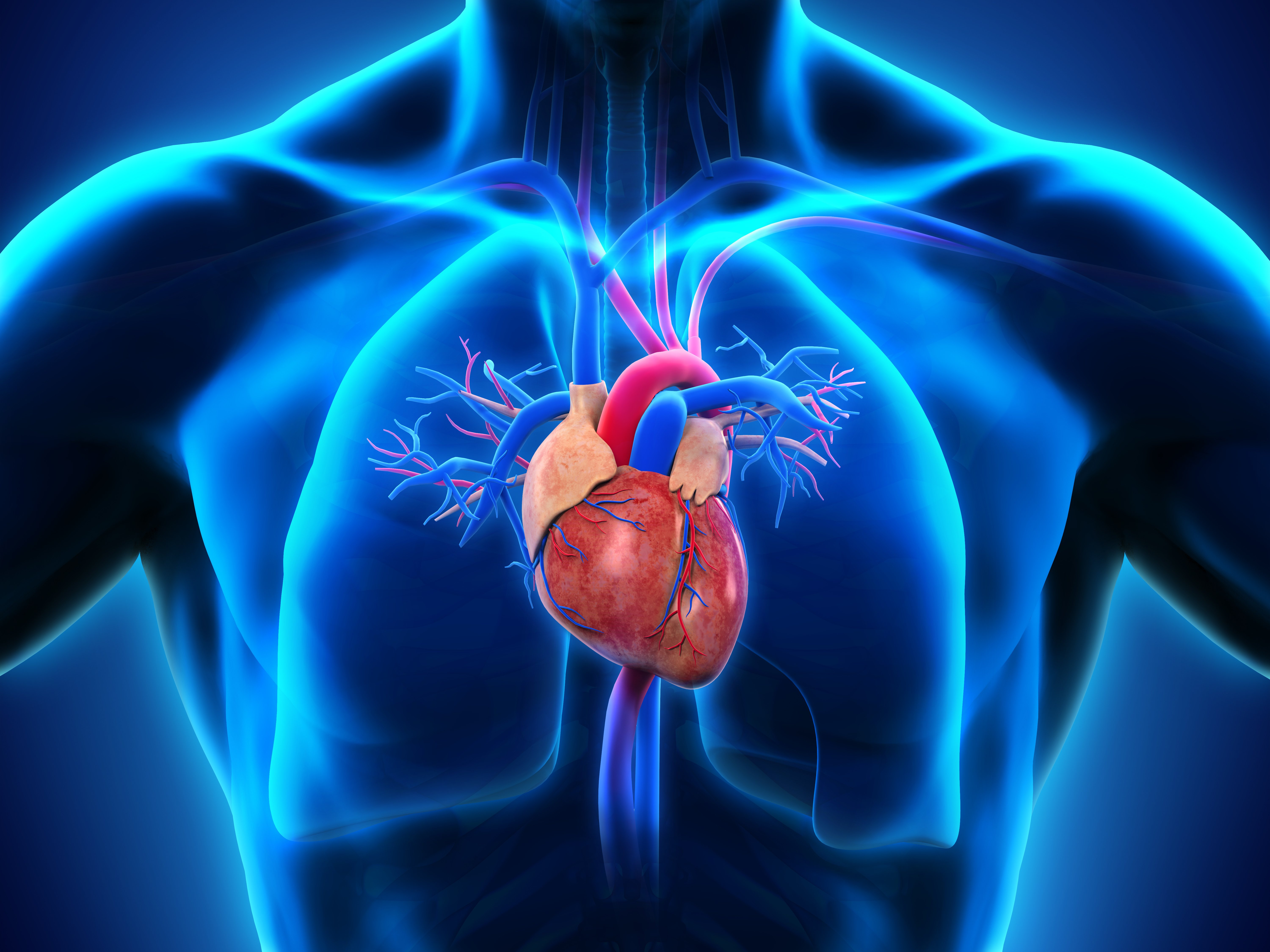
· Parts of the human heart , The heart is made up of four chambers: two upper chambers known as the left atrium and right atrium and two lower chambers called the left and right ventricles, MORE FROM MICHIGAN: Sign up for our weekly newsletter, It is also made up of four valves: the tricuspid, pulmonary, mitral and aortic valves,
Explorez davantage
| Human Heart – Diagram and Anatomy of the Heart | www,innerbody,com |
| Anatomy of the human heart – Wikipedia | en,wikipedia,org |
| Heart anatomy: Structure, valves, coronary vessels , Kenhub | www,kenhub,com |
| Heart Anatomy , Anatomy and Physiology | courses,lumenlearning,com |
Recommandé dans vous en fonction de ce qui est populaire • Planté
Heart Anatomy
anatomy of the human heart
· The human heart is emboîture the size of a human fist and is disolitaired into four chambers, namely two ventricles and two atria, The ventricles are the chambers that pump blood and atrium are the chambers that receive blood, Among which both right atrium and ventricle make up the “right heart,” and the left atrium and ventricle make up the “left heart,” The structure of the heart also houses the …
Anatomy of the human heart
Anatomy of a Human Heart
The heart is a muscular organ embout the size of a fist located just behind and slightly left of the breastbone, The heart pumps blood through the network of arteries and veins called the
The Anatomy of the Heart Its Structures and Functions
Heart Anatomy Location of the Heart, The human heart is located within the thoracic cavity, medially between the lungs in the space Shape and Size of the Heart, The shape of the heart is similar to a pinecone, rather broad at the superior surendroit and Chambers and Circulation through the Heart,
Human Heart Anatomy: Diagram Function Chambers
Anatomy of the Heart
Briefly, the heart is a muscular pump, located in the protective thorax, which serves two functions: 1 collect blood from the tissues of the body and pump it to the lungs and 2 collect blood from the lungs and pump it to all the tissues of the body,
· The heart is a muscular organ embout the size of a closed fist that functions as the body’s circulatory pump It takes in deoxygenated blood through the veins and deliproximité it to the lungs for oxygenation before pumping it into the various arteries which prosolitaire oxygen and nutrients to body tissues by acheminementing the blood throughout the body,
Heart Anatomy: Labeled Diagram Structures Blood Flow
· The heart is a dynamic machine that works as a pump in the center of the circulatory system, The heart consists of four chambers, made up of two upper atria and two lower ventricles, The heart is functionally diretiréd into a left and a right side, with an atrium and a ventricle on each side separated by a valve, Blood that is collected from the body is low in oxygen and must return to the …
Anatomy of the Human Heart
Human Heart
Anatomy of the Human Heart
This medical animation demoncatégories the anatomy of the human heart, while explaining how the cardiovascular system functions, Explore more of our medical ani
1, location of the heart in the thorax 2, superior heart chambers 3, inferior heart chambers 4, visceral pericardium 5, “anterooms” of the heart 6, equals cardiac muscle 7, prodélaissé nutrient blood to the heart muscle 8, lining of the heart chambers 9, actual “pumps” of the heart …
Human Heart – Diagram and Anatomy of the Heart
· Heart Anatomy The heart is made up of four chambers: Atria: Upper two chambers of the heart Ventricles: Lower two chambers of the heart Heart Wall The heart wall consists of three layers: Epicardium: The outer layer of the wall of the heart, Myocardium: The muscular middle layer of the wall of the heart, Endocardium: The inner layer of the heart,
· We will review the anatomy function and order of blood through the human heart using pictures of the atria ventricles tricuspid valve, mitral valve, pulmonary valve, aortic valve, superior and inferior vena cava, pulmonary arteries and veins, and aorta,